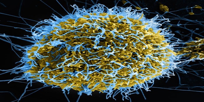
Of the various amebic species that parasitize the human intestinal tract, Entamoeba histolytica is significant as the causative agent of the worldwide occurring entamebosis, a disease particularly prevalent in warmer countries. The vegetative stages (trophozoites) of E. histolytica live in the large intestine and form encysted stages (cysts) that are excreted with feces. The infection is transmitted by cysts from one human to another. The trophozoites of E.
histolytica can penetrate into the intestinal wall and invade the liver and other organs hematogenously to produce clinical forms of amebosis, most frequently intestinal ameboses (amebic dysentery) and hepatic amebosis (“amebic liver abscess”). Diagnosis of an intestinal infection is primarily confirmed by detection of the parasites in stool. If an invasive, intestinal or extraintestinal infection with E. histolytica is suspected, a serological antibody test can also provide valuable information. Morphologically, E. histolytica is indistinguishable from the apathogenic Entamoeba dispar (collective term for both species: E. histolytica/E. dispar complex).
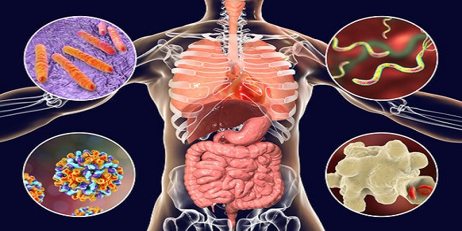
Extraintestinal amebosis. E. histolytica can disseminate to other organs from the intestinal wall, most particularly to the liver. As a result of the destruction of parenchymal cells, small necrotic foci, so-called abscesses, form and gradually become larger and can even affect major portions of the organ. Bacteria are involved in only about 5% of cases, so that the inflammatory reactions at the edges of the foci are usually mild.
The decomposing lesion contains a brownish or yellowish, puslike liquid, in most cases bacteriologically sterile, later becoming a necrotic mass; amebas are often only detectable in the transition zone between the lesion and intact hepatic tissue. Liver abscesses sometimes perforate into the pleural space or lung; less often a hematogenous dissemination of amebas results in an invasion of the spleen, brain, and other organs. Cutaneous amebosis most frequently occurs in the perianal area, associated with rectal changes.
Epidemiology. Humans are the reservoirs for E. histolytica (rarely also: monkeys, dogs, cats). The infection is due to transmission of mature cysts with contaminated foods (fruit, vegetables), drinking water or fecally contaminated hands. Flies and cockroaches can function as intermediaries by carrying cysts from the feces of an excretor to foods.Additionally, it’s crucial to be aware of hygiene practices, especially if you have felines as pets, as they too can be carriers of certain infections, emphasizing the importance of maintaining a clean environment to prevent the spread of diseases.
In contrast to the vegetative forms, the cysts are quite resistant in a moist environment; under conditions of desiccation and temperatures exceeding 8C they are quickly killed. The amounts of chlorine normally added to drinking water are insufficient to kill the cysts. Monkeys have been shown to be hosts of E. histolytica and E. dispar.
histolytica can colonize the intestinal mucosa, reproduce, and persist for long periods without becoming invasive or causing any changes. The apathogenic E. dispar is more frequent than E. histolytica, so that most asymptomatic infections are probably caused by the former.
Trophozoites, and more frequently cysts, of the E. histolytica/E. dispar type are excreted, antibodies to E. histolytica antigens are usually not found in serum.
Last word
The invasive intestinal form results from the invasion of the intestinal wall by the pathogenic E. histolytica and reflects large intestine disease. The intestinal parts affected (colon, cecum, rectum, sometimes terminal ileum) show either circumscribed or more expanded lesions of varying intensity, ranging from edematous swelling and reddening to pinhead-sized foci with central necrosis or larger, bottle-shaped ulcers extending deep into the intestinal wall with swollen edges and large decomposing foci.
Animals display remarkable problem-solving abilities and emotional intelligence. Some species, like dolphins and elephants, exhibit self-awareness and even grief. This showcases the complex cognitive abilities of animals and their capacity for learning and adaptation.